A new study provides the most detailed look at the brain region that controls movement and could help fight diseases like ALS and dementia
SEATTLE, WA, UNITED STATES, September 23, 2026 /EINPresswire.com/ — The brain’s motor cortex plans, controls, and executes voluntary muscle movements. Whether you’re playing catch, writing your name, or chewing food, it all points back to this important region and its connections to other parts of the brain.
Yet despite extensive study, scientists still haven’t created a detailed map of how the mouse motor cortex is internally organized. Most brain atlases have divided it into two general regions—primary and secondary—based on differences in connectivity, but this doesn’t provide a detailed or consistent picture.
A new study by researchers at the University of Basel, Friedrich Miescher Institute for Biomedical Research, and the Allen Institute paints a more precise portrait of the mouse motor cortex. Scientists mapped this critical area into 16 distinct subregions, each with its own pattern of connections. Their results could have important implications for understanding diseases that affect the motor cortex, such as amyotrophic lateral sclerosis (ALS) and frontotemporal dementia (FTD).
“Understanding the precise wiring of the brain is essential for developing effective treatments for brain diseases. Here, our Swiss collaborators’ expert charting of the functionally specific motor cortex circuit, combined with the Allen Institute’s foundational connectivity atlas, resulted in such a precision map that drives movement control,” says Hongkui Zeng, executive vice president and director of Brain Science at the Allen Institute and a senior author on the study.
[Motor cortex’s geography in unprecedented detail]
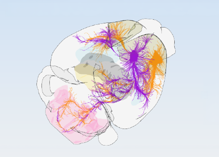
Researchers studied 547 experiments that tracked how nerve signals travel out of the motor cortex and into other parts of the brain. They looked at where each tiny section of the motor cortex sends its “messages”—to regions involved in movement, sensory information, and decision-making.
By grouping areas that send similar signals, they found 16 distinct subregions, each responsible for something slightly different. These areas are arranged in three rows, organized in two main ways:
1. Moving from back to front separates areas connected to sensory information (like touch and body position) from areas connected to planning and decision-making.
2. Moving sideways reveals areas linked to different body parts—from the trunk and limbs on one side, to the face and mouth on the other.
Researchers also confirmed their map using two other methods: tracing individual neurons and looking at what types of brain cells were present. All three methods pointed to the same 16 subregion structure.
Additionally, scientists discovered that the primary motor cortex and secondary motor cortex work in tandem as they send signals to the brainstem and spinal cord, which challenges previous assumptions that they were arranged in more of a hierarchy.
“The most fascinating finding is the extremely high precision with which the motor cortical modules interact with the output regions and that the modules communicate to the rest of the cortex using the same wiring logic,” says Silvia Arber, professor of neurobiology at the University of Basel and the Friedrich Miescher Institute for Biomedical Research, and lead author of the study.
Scientists used the Allen Institute’s Mouse Brain Atlas for their research to leverage the anatomical databases and workflows needed for this high-precision work. The map is also available for researchers around the world through an online tool called BrainGlobe, which allows scientists to work from the same reference point.
“Researchers interested in the cortex now have an accessible unified map to align their data to, and this will accelerate progress in the field,” says Antonio Falasconi, one of the study’s first authors. “Bringing together vast datasets describing the brain’s wiring and its cellular makeup, we discovered a valuable and much more precise underlying blueprint of motor cortex organization,” says Harsh Kanodia, another first author of this study.
[Practical implications]
Amyotrophic lateral sclerosis (ALS) and frontotemporal dementia (FTD) both affect the motor cortex—ALS by destroying cells that send signals to the muscles, while FTD damages the frontal part of the brain that dictates personality, behavior, and language. However, scientists don’t fully understand why some cells are targeted and others are not. This new detailed map could change that. Scientists can now look at which of the 16 subregions has vulnerable cells, while also tracking how disease spreads through connected regions. Future research could also look into how the 16-subregion system relates to the cortical organization in other species, including humans.
Peter Kim
Allen Institute
+1 206-605-9884
email us here
Legal Disclaimer:
EIN Presswire provides this news content “as is” without warranty of any kind. We do not accept any responsibility or liability
for the accuracy, content, images, videos, licenses, completeness, legality, or reliability of the information contained in this
article. If you have any complaints or copyright issues related to this article, kindly contact the author above.

Media gallery